If your vet suggests “doing some diagnostic imaging” they will most commonly mean taking radiographs (x-rays) or performing ultrasound scans. See below for a handy guide as to what these are used for and how they can help to diagnose your pet.
Radiography (taking x-rays)
People commonly associate radiographs with looking for broken bones, but they are also useful for looking at other parts of the body such as the thorax (chest) and abdomen (tummy). It is important that the animal is still for radiography, therefore it will often require using a sedation or general anaesthetic (when your pet is fully asleep) to allow them to get the best pictures.
How it works
X-rays are emitted from a special machine and pass through the body to create an image on a film or plate (the radiograph) in a similar way to how light does for a photograph. Denser (heavier) parts of the body stop more x-rays passing through, which means that the picture created allows us to distinguish areas of different density. Bone and teeth are the most dense and appear white, softer tissues (organs such as the liver and muscles) and fluids appear grey, and air (such as that in the lungs) appears black. Metal will show up as a very bright white so we can see things like the implants used to fix broken legs, or foreign bodies if your pet has eaten something other than food!
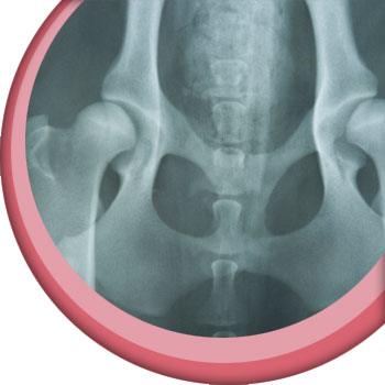
What it’s used for
• Bones (known as orthopaedics) – x-rays are useful to look for broken bones as well as tumours of bones and joint problems like osteoarthritis (inflammation in or around a joint).
• Tummy (abdomen) – your vet can use x-rays to look at the size and position of organs, as well as to look for any foreign bodies in the intestines although only some of these will show up. They can also see some tumours and some types of bladder stones.
• Chest (thorax) – your vet can look at the size and position of the heart, whether there is any air or fluid on or around the chest where it shouldn’t be, and whether there are any tumours. They can also look for any areas of the lungs that look abnormal.
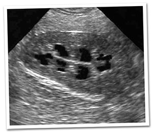
Ultrasound scans
An ultrasound scan is the same as that used for pregnancy scans in people.
It does not require the animal to be as still as for x-rays, therefore they can
usually be done with your pet awake or under a low level of sedation,
unless they are particularly wriggly!
How it works
A probe is placed on the skin which gives off ultrasound waves. These bounce back off the
organs in the body in different ways which allows a picture to be created by the machine.
What it’s used for
• Pregnancy diagnosis – an ultrasound scan can confirm the presence of fetuses (puppies or kittens before they are born) in the uterus (womb) but cannot be used to give reliable numbers as they can look like a bit of a jumble.
• Heart scans – the blood flowing through the heart and the way the heart muscle is moving can be assessed. This allows your vet to judge how well the heart is working. These are often done by specialist vets.
• Abdominal scans – these allow your vet to look at the organs within the abdomen including the liver, stomach, intestines, bladder and kidneys. In male dogs they can look at the prostate gland. An ultrasound scan gives a better idea of the 3D structure than an x-ray, but depending on what they are looking for, your vet will often do both procedures to be able to get the most information and decide on the best treatment.
MRI/CT scans
Some specialist referral vet practices have MRI (magnetic resonance imaging) or CT (computed tomography) scanners. Mobile MRI scanners also exist that may visit different practices. These are more advanced methods of imaging that can be very useful to look at the brain and spine in pets with neurological conditions (problems with the nerves in the body, be it in the brain, spine or elsewhere). Animals need to be still for these procedures, particularly for MRI, therefore general anaesthesia is required.
CT uses x-rays to build up a picture of an area of the body. It is very good for looking at bones. MRI scans use magnetic and radio waves to build up a picture. They are good at producing detailed images of soft tissues, such as the brain, spinal cord and internal organs.
Disclaimer: This website has been designed to offer information surrounding the use of antibiotics and infection control for pet owners. It does not replace advice from your veterinary surgeon. If you believe your pet is unwell or you have any questions relating to their treatment, please always contact your veterinary surgeon for advice.