Cytology
Cytology means the study of cells. Cells are the building blocks that make up our bodies, for example red blood cells which carry oxygen or skin cells which protect us from the outside world. There are many different types of cells all with their own job within the body. They can only be seen using a microscope, but looking at which type of cells are present in a sample, and their appearance, can give clues as to what disease your pet may have.
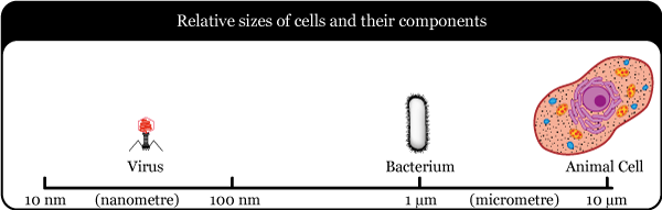
We can also look for bugs that may be present, for example bacterial or yeast cells. Bacteria only have one cell and it is much smaller than an animal or human cell. Viruses are much too small to be seen with most microscopes, and require special techniques only available at certain laboratories, such as those in universities.

There are a few different ways of obtaining samples for cytology which your vet may use:
• “Impression smears” from skin – the microscope slide is pressed directly onto the affected area of skin to pick up cells from the surface.
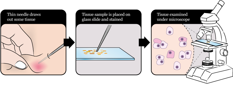
• “Fine needle aspirates” – this is a technique where a needle is inserted into a lump and a small amount of fluid or material is collected.
• Fluid collected from the airways.
• Swabs (like long cotton buds) of any bodily discharges, for example from the nose, ears or eyes.
To carry out cytology on a sample your vet or the laboratory will make a smear on a microscope slide creating a thin coating of sample material. Once this is dry they will dip it in a special stain that allows cells to be seen more clearly.
What your vet is looking for:
• Inflammatory cells – these are also called white blood cells and they are part of the immune system. They appear when there is inflammation or injury to part of the body. How many and what type they are may be helpful for determining the type of disease process.
• Bugs – bacteria, yeasts or fungi. The shape of bacteria may be important to determine the type of infection and which antibiotics are more likely to be successful.
• Tumour cells – the type of cell affected and their appearance can give an idea as to whether the tumour is likely to be harmless (benign) or harmful (malignant – which are often more likely to spread and cause problems).
For skin disease your vet may also take skin scrapes and hair plucks to look for parasites under the microscope. A skin scrape involves using a small blade to scrape cells and debris off a section of skin. As many mites burrow down into the skin, this is the best way to try to find them, but as they are good at hiding just because we don’t see them doesn’t mean they’re not there! As this can occasionally be a little bit uncomfortable for your pet, sedation may be recommended by your vet.
Hair plucks are exactly as they sound – the vet will pluck out a few hairs and then examine them directly under the microscope. As most pets usually have a few hairs to spare, sedation is not usually necessary for this!
Disclaimer: This website has been designed to offer information surrounding the use of antibiotics and infection control for pet owners. It does not replace advice from your veterinary surgeon. If you believe your pet is unwell or you have any questions relating to their treatment, please always contact your veterinary surgeon for advice.